


The economic benefits of sheep farms are directly related to the reproductive characteristics of sheep, and veterinary ultrasound plays a very important role in the diagnosis of pregnancy in ewes. The pregnancy of the ewe can be determined by ultrasound examination.
The breeder/veterinarian can analyze the results of ultrasound detection and carry out scientific feeding of pregnant ewes by grouping and individual housing, so as to improve the nutritional management level of pregnant ewes and increase the lambing rate.
At this stage, the more commonly used method for checking ewe pregnancy is to use veterinary B/W ultrasound machine.
Veterinary B/W ultrasound is usually used for pregnancy diagnosis, disease diagnosis, estimation of litter size, identification of stillbirths, etc. It has the advantages of rapid examination and obvious results. Compared to traditional testing methods, veterinary ultrasound greatly simplifies the inspection process, reduces inspection costs, and allows the owner/veterinarian to quickly identify problems and take quicker action, e.g., rapid herd separation.
What is B/W ultrasound?
B/W ultrasound is a high-tech means to observe the living body in cross-section without any damage or stimulation. It has become a favorable assistant for veterinary diagnostic activities and an essential monitoring instrument for scientific research such as live egg collection and embryo transfer.

B/W ultrasonic probe probing site and method
(1) Probing site
In the early stage of pregnancy, the probe can be carried out on both sides of the breast and the less hairy area between the breasts, or the interval between the two breasts. In the middle and late stages of pregnancy, the right abdominal wall can be explored. There is no need to cut the hair in the less hairy area, but the side abdominal wall needs to be cut, and the rectal exploration needs to be done in the right side of the breast.
(2) Probing method
The probing method is basically the same as that for pigs, the examiner squats on the side of the sheep body, and after applying coupling agent locally or on the probe, the probe is pressed against the skin towards the direction of pelvic inlet to carry out a fixed-point fan-shaped sweep. Sweeping from the breast straight forward to backward, from both sides of the breast to the center, or from the center of the breast to both sides is acceptable. Early in pregnancy, the sac is not large and the embryo is small enough to require a slow sweep to detect it. The inspector can also squat behind the rump of the goat and hold the probe from the middle of the two hind limbs of the goat to the udder for scanning. If the udder of dairy goat is too large, or the hair on the lateral abdominal wall is too long, which affects the visibility of the probing site, the assistant can lift up the hind limb on the probing side, exposing the probing site, but it is not necessary to cut the hair.
B/W ultrasound examination of ewes in a holding position
Generally, the ewe should stand naturally, and the assistant should support her and keep quiet, or the assistant should clamp the neck of the ewe with both legs to keep her in place, or a simple holding frame can be used to keep the ewe in place. Side-lying bailing can slightly advance the diagnostic date and improve the diagnostic accuracy, but it is not convenient to use in a large group.B/W ultrasound can be used to detect early pregnancy in side-lying, supine or standing position.
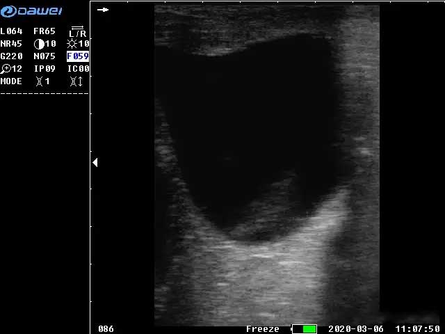
In order to make a good distinction between artifacts, we must recognize a few typical B/W ultrasound hyperimages in sheep.
(1) Ultrasound image characteristics of ewe follicles on sheep B/W ultrasound:
From the shape, most of them are round, and a few of them are oval and pear-shaped; from the echo intensity of sheep B images, because the follicle is filled with follicular fluid, sheep B/W ultrasound scans are echo-less, and they are a dark area on the image, which is a clear contrast to the strong echogenic (bright) area of the follicle wall and surrounding tissues.
(2) Sheep B/W ultrasound imaging features of the corpus luteum:
Shape-wise most of the corpus luteum tissue was round or ovoid. Since the luteal body tissue is weakly echogenic on ultrasound scanning, it is not as dark as that of the follicle from the color of the sheep B/W ultrasound image. In addition, the biggest difference between the ovary and the corpus luteum on sheep B/W ultrasound image is that there are trabeculae and blood vessels in the luteal body tissue, so that there are scattered dots and lines of bright areas on the imaging, whereas there are none in the follicle.After the examination, the examined sheep are marked and then divided into flocks.
Precautions for practical application
1. Do not use the instrument in close proximity to equipment such as generators, dentistry, radio, etc., as this may interfere with the image and affect the quality of the image.2. Before using the instrument for the first time, you must read the instruction manual of B/W ultrasound carefully and operate it strictly according to the requirements.
3. In the process of operation, do not pull down the probe, to protect its surface, you can rub the coupling agent on the surface of the probe, so as to make good contact between the examined animal and the probe.
4. Practice more to skill the operation process and check it quickly.
5. Be gentle with the sheep grasping work, not violent grasping, otherwise it may lead to abortion and other problems of the sheep.
Post time: Jul-04-2024