


Renal ultrasound in pet dogs requires a 5MHz or 7MHz probe. Gas can be disruptive to the results of the sweep; minimize gas in the intestines before the sweep. The skin is cleaned by shaving the last two intercostals on the right side and behind the last rib on the left side; the probe is coated with coupling agent so that the probe fits snugly against the skin; and the animal is allowed to relax as much as possible during the sweep to avoid excessive inhalation, which can increase the amount of gas in the bowel. The left kidney is swept behind the quaternary ribs, and the right kidney is swept between the last two ribs or behind the quaternary ribs.
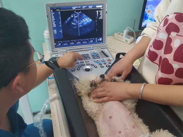
Pet Renal Ultrasound
Perfect function, configuration
Suitable for pet abdominal, obstetrical, cardiac, vascular, small organ and urological examinations
- Main screen 15-inch high-resolution color LCD monitor;
- With professional probe placement rack ≥ 2;
- USB3.0 interface clinical picture, image video, report storage, export;
- Built-in probe interface ≥ 2, full activation of the same size, interoperability and interoperability;
- Movie playback ≥ 3000 frames, support manual and automatic playback;
- With professional probe placement rack ≥ 2;
- Configuration of ultrasound graphic workstation set;
- Hold a foot switch;
- Digital hard disk capacity ≥ 256G;
Flexible adjustment within 45° of the display
Full consideration of ergonomic design, maximize to meet the needs of doctors with different perspectives, reduce visual fatigue.
Excellent system functions to meet the needs of multi-disciplinary diagnosis
With more sensitive echo frequency shift capture capability, thus obtaining better contrast resolution, more tissue structure information, and clearer and more detailed clinical images.
Equipped with high-end imaging technology, providing comprehensive diagnosis and treatment programs for clinical medical treatment.
Powerful image processing functions, high-end technology applications: harmonic imaging, tissue Doppler imaging, elasticity imaging, contrast imaging, Pview wide-view imaging technology, and so on.
Superior Image Processing Functions
Digital beam enhancer
Multiple beam synthesis
Anatomical M-mode with ≥ 3 sampling lines
Doppler imaging
Spectral Doppler imaging
Spatial composite imaging
Frequency composite imaging
Extended imaging
Post time: Jul-25-2024