In the reproductive process of sows, one of the primary concerns for breeders is whether the mating has been successful. To address this issue, the most convenient method is to use ultrasound for examination. Ultrasound technology, with its non-invasive, real-time, and accurate characteristics, provides significant support for sow reproduction, helping farmers improve production efficiency and economic benefits.
Veterinary ultrasound detection for pregnancy not only has a high accuracy rate but is also currently the most economical and fastest method available. The principle behind this technology involves the ultrasound probe emitting sound waves into the body’s tissues. As these sound waves penetrate the tissues, they produce varying signals based on the different densities of the tissue media. These signals are then received by the ultrasound probe and returned to the main machine, resulting in imaging. This is how we see different colors; typically, black represents liquid (such as urine or amniotic fluid), while bright white typically indicates denser tissues, such as bone or certain pathological calcifications. Veterinarians can use these images to observe the sow’s reproductive system and assess its physiological status, pregnancy condition, and fetal health.
Applications in Sow Reproduction
Pregnancy Diagnosis
One of the most common applications of ultrasound technology is pregnancy diagnosis. Through ultrasound examination, veterinarians can confirm whether a sow is pregnant at an early stage (usually around 20 days post-mating). This early diagnosis allows farmers to adjust feeding management promptly, thereby improving reproductive efficiency.
Fetal Health Monitoring
With ultrasound technology, veterinarians can monitor the fetuses during pregnancy, assessing their number, size, and health status. This helps in the timely detection of potential pregnancy complications, such as miscarriage or developmental issues in the fetuses, allowing for appropriate interventions.
Reproductive Organ Examination
Ultrasound provides a clear view of the sow’s reproductive organ status, including the health of the ovaries and uterus. This is crucial for evaluating the sow’s reproductive capacity and identifying reproductive system diseases (such as ovarian cysts or uterine inflammation).
Timing of Mating
Ultrasound technology can assist in determining the optimal mating time for sows. By observing the development of follicles in the ovaries, veterinarians can assess the ovulation timing of the sows, thereby increasing the success rate of mating.
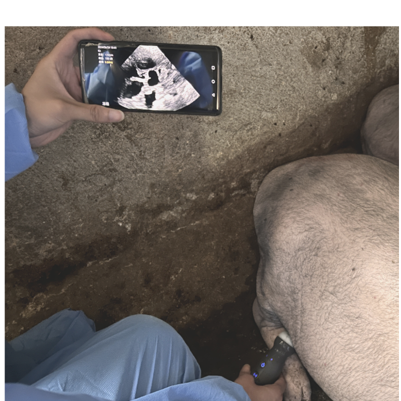
Thus, through different images, it is possible to determine whether mating has been successful. Pregnancy checks can be performed on sows 18 days after mating. In the operation of veterinary ultrasound, the technique and positioning for pregnancy testing are crucial. Only with proficient use and correct positioning can the ultrasound machine’s capabilities be maximized. So, how can one master better pregnancy testing positions? For sows, it is relatively simple. Typically, the testing position is located about five centimeters above the second-to-last and third-to-last pairs of teats. The probe should be directed toward the sow’s spine and moved slowly. Once the image is seen, the veterinarian can determine pregnancy status based on the number of days since mating.
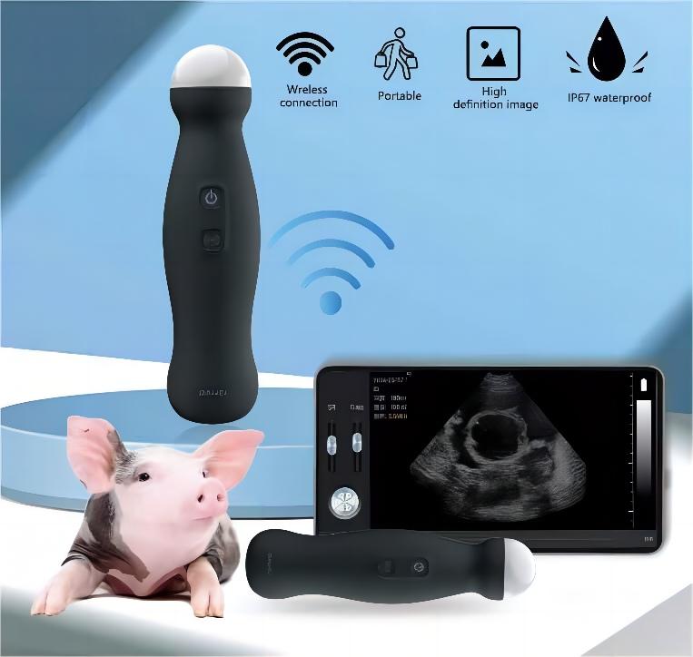
Therefore, monitoring the pregnancy process of sows using ultrasound is very important. A good pig ultrasound machine can provide veterinarians with better diagnostic decisions. Dawi Veterinary Medicine has recently launched a new ultrasound machine for pigs, handheld mechanical wireless ultrasound probe,featuring a 3.5 MHz mechanical sector probe, which is suitable for pigs, sheep, dogs, and cats. This portable and easy-to-use veterinary ultrasound device is designed for real-time diagnosis and comes with a battery and charger.
Post time: Aug-22-2024


