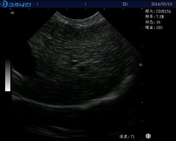
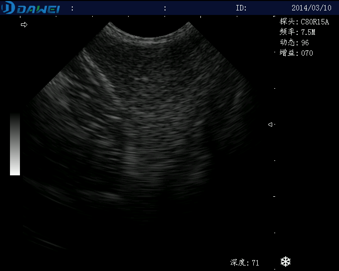
In recent years, veterinary ultrasound has developed rapidly and has become an indispensable diagnostic method in modern clinical medicine. With the in-depth research and wide application of ultrasonic diagnosis in the field of veterinary medicine, abdominal ultrasound has become a valuable auxiliary diagnostic means for abdominal diseases in small animals.
1 General abdominal exploration technique
Ultrasound diagnosis of general abdominal diseases refers to the ultrasound diagnosis of intra-abdominal free fluid, vascular structures, intra-abdominal masses, diaphragm and hernia and other related abdominal diseases. Ultrasound exploration of these abdominal disorders is done in the supine (dorsalrecumbency) position in most cases, and sometimes in the left lateral, right lateral, or standing position. The lower abdomen is clipped, coupling agent is applied, and a 7.5MHz transducer is used for cats and small and medium-sized dogs, while a 5.0MHz transducer is used for large dogs, and a 3.5MHz or 2.5MHz transducer must be used to investigate the deeper structures of the abdomen in large or giant dogs.
2 Liver ultrasound exploration techniques
Intragastric gas is a major obstacle to obtaining high-quality liver images; therefore, diagnostic methods that may cause struggling or aerophagia (gas swallowing) should be avoided prior to ultrasound examination. The animal is held supine on a “V” shaped holding table before the lower abdomen is clipped and couplings are applied. A 5.0 MHz transducer is used for liver ultrasound in most dogs, 3 MHz in large dogs, and 7.5 MHz in cats.
During the examination, the probe is placed firmly under the sternal raphe, and gentle pressure is applied to expel the gas in the stomach in front of the probe. If it is still difficult to see the liver, the examined animal can take a standing, prone (abdominal position) or lateral position – to change the position of the gas in the stomach. The latter two positions can be performed with a plexiglas table (plexiglasstable) with a square hole in the middle, and the scanning is most likely to be successful when performed from below, approaching the abdomen. Fluid from the hypogastric portion of the stomach is used as an acoustic window to aid in the visualization of the liver, and fluid is also helpful in the visualization of the liver if administered to the kidneys.
The entire liver can be explored in both longitudinal and transverse scans, and it is important to ensure that all parts of the liver are visualized when exploring in both dry planes. This usually requires multiple explorations of the entire liver, with the beam directed dorsally and ventrally in the sagittal plane and to the left or right side of the body in the transverse plane, and supplemental visualization of the periphery of the liver with intercostal views, if necessary. A transverse scan in the right lateral 11th or 12th intercostal space is particularly important for visualization of the major abdominal vessels or common bile duct in the vicinity of the hepatic hilum.
3 Ultrasound techniques for the spleen
The spleen is located outside the left circumferential greater curvature of the abdominal cavity. The exact location of the spleen is variable, depending on the degree of gastric dilatation and the size of the other abdominal organs, and the anatomical relationship between the splenic imaging method and the ultrasound image can be clarified in detail under anesthesia. The splenic head (dorsal pole – dorsalextremity> is located below the costal arch, whereas the body and tail (ventral pole – ventralextremity) extend along the left side of the body wall or across the lower abdomen. When the spleen is enlarged it may also extend across the lower abdominal midline or backward into the bladder region. If the spleen cannot be explored from below the side, the head of the sign may be explored on the left side at the 11th or 12th intercostal space. The remainder of the spleen is scanned in a longitudinal or transverse system on the lower abdominal wall. Because of the superficial location of the spleen, scanning with a 7.5 MHz. 10.0 MHz transducer in dogs and cats provides high-resolution images. If the body wall surface nearfield images cannot be fully visualized . The use of a padding block helps to position the spleen away from the transducer while please chu display.
The splenic vessels consist of the splenic artery and the splenic vein: the branches of the splenic vein are clearly visible near the splenic hilum, and the splenic vein often extends a considerable distance to the portal vein, but is sometimes difficult to visualize completely because of the obstruction of intestinal gases. If necessary, Doppler ultrasonography can help to localize them.
4 Ultrasoundof the pancreas
Pancreatic ultrasonography can be taken supine, prone, lateral or standing position, usually, the sound and image workers in dogs or cats to take supine from the lower abdomen, however, when the intestinal lumen there is excessive gas, but also to take the left lateral recumbent position for exploration, should be removed from the left side of the 11 or 12 help between the coat in order to facilitate the propagation of sound, in order to avoid the influence of intestinal gas, can be taken in prone position or lateral recumbent position for exploration, and can be used with the table surface of the plexiglass table perforation from below for exploration. A plexiglass table with holes in the surface can be used for exploration from below. Probing from below facilitates the upward movement of intestinal gas. Pancreatic exploration in dogs can be performed with a 3.0MHz or 7.5MHz transducer, but large dogs need a 3.0MHz transducer and cats need a 7.5-10.0MHz transducer. If it is difficult to visualize the near field in the right side of the body, the use of a cushioning block can be helpful, especially in cats, and in medicine, filling the stomach with water or saline can provide a good acoustic window for the pancreas, which is also used to diagnose the stomach and the skin in animals. However, this technique is not universally recognized in animals because it can induce vomiting, introduce gas and cause additional pancreatic enzyme release. Preliminary studies have also concluded that intragastric instillation of fluid in healthy dogs does not provide a significant acoustic window for ultrasonographic exploration of the pancreas, and that diagnostic ascites can also enhance pancreatic visualization when other noninvasive methods fail, but this method cannot be used routinely in clinical practice, and that an overflow of warmed Ringer’s solution containing sodium chloride or lactate (approximately 60 mL/kg) can be delivered into the posterior peritoneal cavity or into the right umbilicus. The choice of fluid administered is determined by the electrolyte disturbance present in the affected animal. Diseases with impaired regulation of extracellular fluids, such as progressive cirrhosis, congestive heart failure, oliguric renal failure, and severe hemoglobinopenia, contraindicate the use of ascites to assist in ultrasound exploration.
Post time: Dec-12-2023


