


The adrenal glands are vital endocrine organs in dogs, responsible for secreting various hormones such as adrenaline and cortisol. These hormones play crucial roles in a dog’s metabolism, immunity, and stress responses. When adrenal gland function is compromised, it can severely impact a dog’s health. Therefore, timely and accurate diagnosis of adrenal gland diseases is essential. Ultrasound scanning is a non-invasive and painless diagnostic tool widely used in veterinary practice. This article will introduce the ultrasound scanning methods for canine adrenal glands.
Preparation for Canine Adrenal Gland Ultrasound Scanning
Before performing an ultrasound scan, several preparatory steps are necessary to ensure the accuracy and effectiveness of the examination:
- Fasting and Withholding Water: To avoid interference from gastrointestinal contents, it is usually recommended to fast the dog for 8-12 hours before the examination.
- Shaving and Cleaning: To obtain clear ultrasound images, the hair in the examination area should be shaved, and the skin should be cleaned with an appropriate cleaning agent.
- Calming the Dog: Keeping the dog calm and relaxed is very important. Gentle words and petting can help soothe the dog, and mild sedatives can be used if necessary.
- Positioning: The dog is typically placed in a supine position, but depending on the specific situation, lateral or standing positions may also be used. Ensure the dog remains still throughout the examination.
- Probe Selection: Choose a probe with an appropriate frequency, generally using a high-frequency probe (7.5-12 MHz), as it provides high resolution suitable for detailed observation of the adrenal glands.
- Probe Placement: Place the probe on the dog’s abdomen, slightly towards the lateral side. According to anatomical positioning, the adrenal glands are located anteromedial to the kidneys.
- Parameter Adjustment: Adjust the ultrasound machine’s parameters, such as gain and depth, based on the dog’s size and specific conditions to obtain the best image.
Steps for Canine Adrenal Gland Ultrasound Scanning
- Positioning: The dog is typically placed in a supine position, but depending on the specific situation, lateral or standing positions may also be used. Ensure the dog remains still throughout the examination.
- Probe Selection: Choose a probe with an appropriate frequency, generally using a high-frequency probe (7.5-12 MHz), as it provides high resolution suitable for detailed observation of the adrenal glands.
- Probe Placement: Place the probe on the dog’s abdomen, slightly towards the lateral side. According to anatomical positioning, the adrenal glands are located anteromedial to the kidneys.
- Parameter Adjustment: Adjust the ultrasound machine’s parameters, such as gain and depth, based on the dog’s size and specific conditions to obtain the best image.
Techniques for Canine Adrenal Gland Ultrasound Scanning
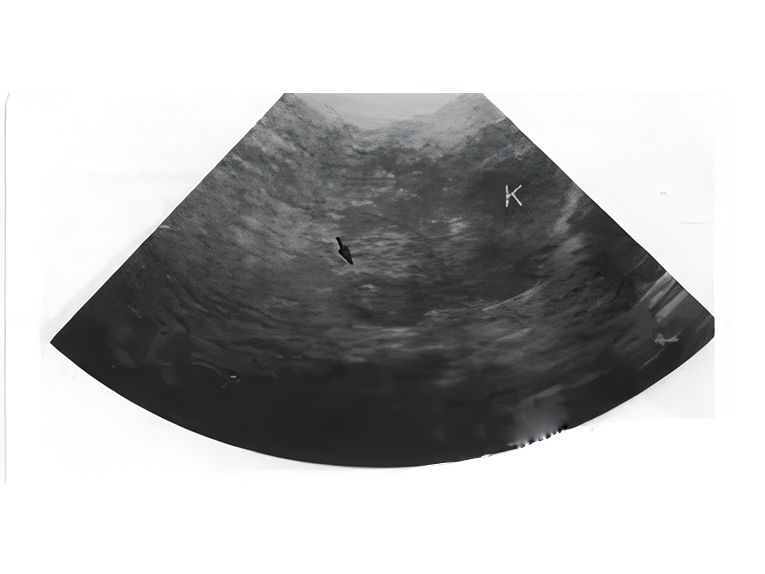
- Longitudinal Scanning: Scan longitudinally along the midline of the abdomen to observe the long axis of the adrenal glands. Adrenal glands typically appear elongated or oval, with a smooth surface and uniform echogenicity.
- Transverse Scanning: After longitudinal scanning, perform transverse scanning to comprehensively observe the shape and size of the adrenal glands. Transverse scanning helps identify the cross-sectional structure of the adrenal glands.
- Comparative Observation: The adrenal glands are usually located anteromedial to the kidneys, with a noticeable echogenicity contrast with the surrounding tissues. Normal adrenal glands have echogenicity slightly higher than the kidneys but lower than the surrounding fat tissue.
- Dynamic Observation: Through dynamic observation, the blood flow and functional status of the adrenal glands can be evaluated. Doppler ultrasound can help identify the vascular structure and hemodynamic changes of the adrenal glands.
Common Problems and Solutions
- Blurry Images: This could be due to poor probe contact or insufficient ultrasound gel. Reposition the probe and increase the amount of ultrasound gel.
- Difficulty Locating Adrenal Glands: The adrenal glands are small and deeply located, and multiple attempts from different angles and positions may be needed. Anatomical landmarks (such as the kidneys and liver) can aid in localization.
- Interference Echoes: Gastrointestinal gas, fat tissue, and other factors may produce interference echoes. Fasting, withholding water, and appropriate adjustment of ultrasound parameters can reduce interference.
Conclusion
Ultrasound scanning of the adrenal glands is an important tool for diagnosing adrenal gland diseases in dogs. With proper preparation and accurate scanning techniques, clear ultrasound images can be obtained, aiding veterinarians in precise diagnosis and treatment. Timely detection and treatment of adrenal gland diseases are crucial for ensuring the health of dogs. It is hoped that the methods and techniques provided in this article will be helpful in veterinary clinical practice.
Post time: Aug-08-2024