


Generally pet ultrasound images, mainly from the following dimensions:
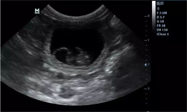
1, pet ultrasound imaging if black, indicates the detection of low-density liquid substances, such as urine in the bladder, amniotic fluid in the uterus, follicular fluid in the follicle, blood in the pelvic wall blood vessels, etc.
2. A white ultrasound image indicates the detection of high-density organs or tissues, such as growths in the musculoskeletal layer.
3. A gray pet ultrasound image indicates the detection of medium density tissue or organs, such as muscle tissue in the uterine horns, fetal muscle tissue, and so on.
So when we use veterinary ultrasound, to the dog to do the examination process need to do supine bailing work, when the stomach is empty when the dog’s stomach wall on the veterinary ultrasound showing alternating layers of high and low echoes, the thickness of the stomach wall is 3-5 millimeters, the duodenal wall thickness is 2-3 millimeters, the contents of the gastrointestinal tract liquid is not echoey, the gas is obviously high-intensity echoey interfaces, normal peristaltic wave.
The dog’s pancreas for veterinary ultrasound sonograms need to be supine, lateral recumbency for fasting time for sweeping, need to be in the 1-2 lumbar vertebrae plane line transverse sweep abdomen, for up and down movement, the right low and left high position oblique for sweeping, the dog’s pancreas image long-axis right low and left high present oblique shape, the border is neat and smooth, the dog’s pancreas internal echogenicity present tiny light spots, the surface is cleaned and leveled, the echogenicity level is not lower than that of the liver of the animals. The echoes of the pancreas were elevated in obese and old dogs.
Post time: May-15-2024