Veterinary ultrasound machines are most commonly used in the ultrasound examination of canines and small animals for abdominal and thoracic ultrasound. Each of these exams has its own objectives, but are complementary when used together in a patient, and it is often recommended that all three exams be completed in a test.
Veterinary ultrasound machines with microconvex probes are generally used for ultrasound examination of small animals such as dogs, with common frequency settings ranging from 5 MHz for large dogs (> 20 kg) to 7 MHz for small dogs (dogs and cats ≤ 20 kg).
Preparation for testing with a veterinary ultrasound machine:
Detection is usually done without shaving, by separating the fur and applying the couplant to the desired area. However, sometimes when a situation is encountered where the image quality is poor due to thicker hair in the dog and more detail is needed, shaving can improve the image quality.
When applying the couplant, it is necessary to prevent the formation of air bubbles (especially if there is no shaving, as the couplant may develop air bubbles in the fur, which can affect the image quality).
Specific scanning technical points for canine detection with a veterinary ultrasound machine:
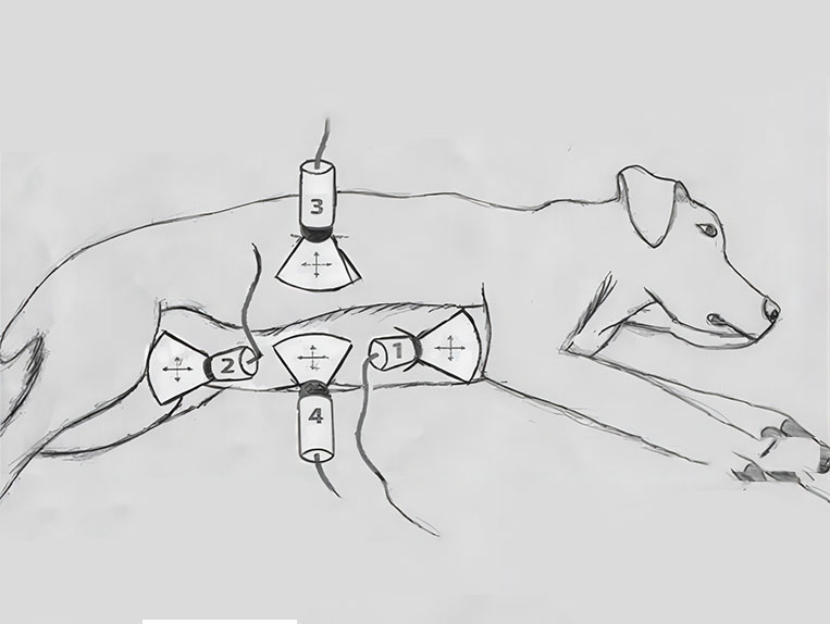
A systematic four-view examination is required when the dog is in right or left lateral recumbency (the most common position) or sternal (with the hind legs turned toward the sonographer to allow access to the subxiphoid and bladder areas) or standing (see figure below). The 4 sites of assessment are included:
To perform the ultrasound, the patient can be placed in the left or right lateral recumbent position, or the sternum can be scanned. This image shows the left lateral recumbent position. The 4 sites to be evaluated include the subxiphoid or diaphragmatic muscle (DH) site (1), the right parietal lumbar or hepatic and renal (HR) site (2), the bladder colic (CC) site located above the midline of the bladder (3) and the left parietal lumbar or splenic and renal (SR) site (4). If the aim is to identify free fluid, a variant of the technique can be substituted, so that instead of a gravity-dependent view a “flash” scan can be performed (position 4 for dogs in left lateral recumbency or position 3 for dogs in right lateral recumbency) and renal assessment is not important. At each site, the ultrasound probe was initially placed longitudinally over the underlying organs and spread out in a fan-like pattern at a 45° angle and moved 2.5 cm in cranial, caudal, left, and right directions.
Important considerations for testing dogs with a veterinary ultrasound machine
Patients with a negative scan on veterinary ultrasonography and stable symptoms or persistent clinical signs usually benefit from serial examinations.
A negative veterinary ultrasonography scan cannot rule out internal damage or pathology. Ultrasonography is unlikely to detect pathology that is located more than a few millimeters inside the lungs and does not extend to the periphery. Veterinary ultrasonography is also structurally specific and focused, and pathology may be missed in areas not evaluated within the unscanned area.
Patients with asthma or shallow breathing may be difficult to assess for signs of gliding if B-lines are not present.
Gliding signals are visible only during the dynamic phases of inspiration and expiration and disappear between breaths (during the static phase of respiration and during apnea). Single-lung intubation (i.e., left or right mainstem bronchial intubation) will result in a lack of gliding signs in the unintubated lung.
Movement of your hand, the probe, or the patient may cause false-positive glide signs; keep your hand, the patient, and the ultrasound probe still when looking for glide signs. Abdominal scans may produce false-positive results when normal hypoechoic abdominal structures are interpreted as free body fluids. Structures commonly misinterpreted as free fluid include the gallbladder, common bile duct, hepatic veins, caudal vena cava, and occasionally the gastrointestinal tract wall and/or gastrointestinal tract contents. Using both transverse and longitudinal views helps to avoid misinterpretation of normal abdominal structures.
Changing the depth and focus of the ultrasound window may help with the sensitivity of recognizing small volumes of fluid during abdominal scanning examinations.
Post time: Jun-08-2024


